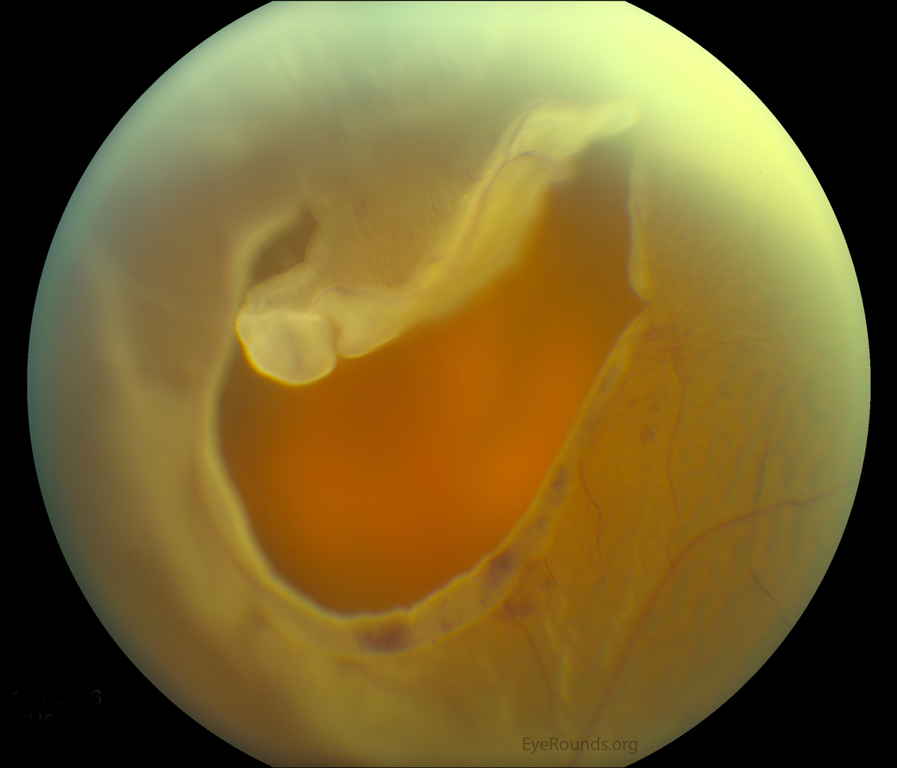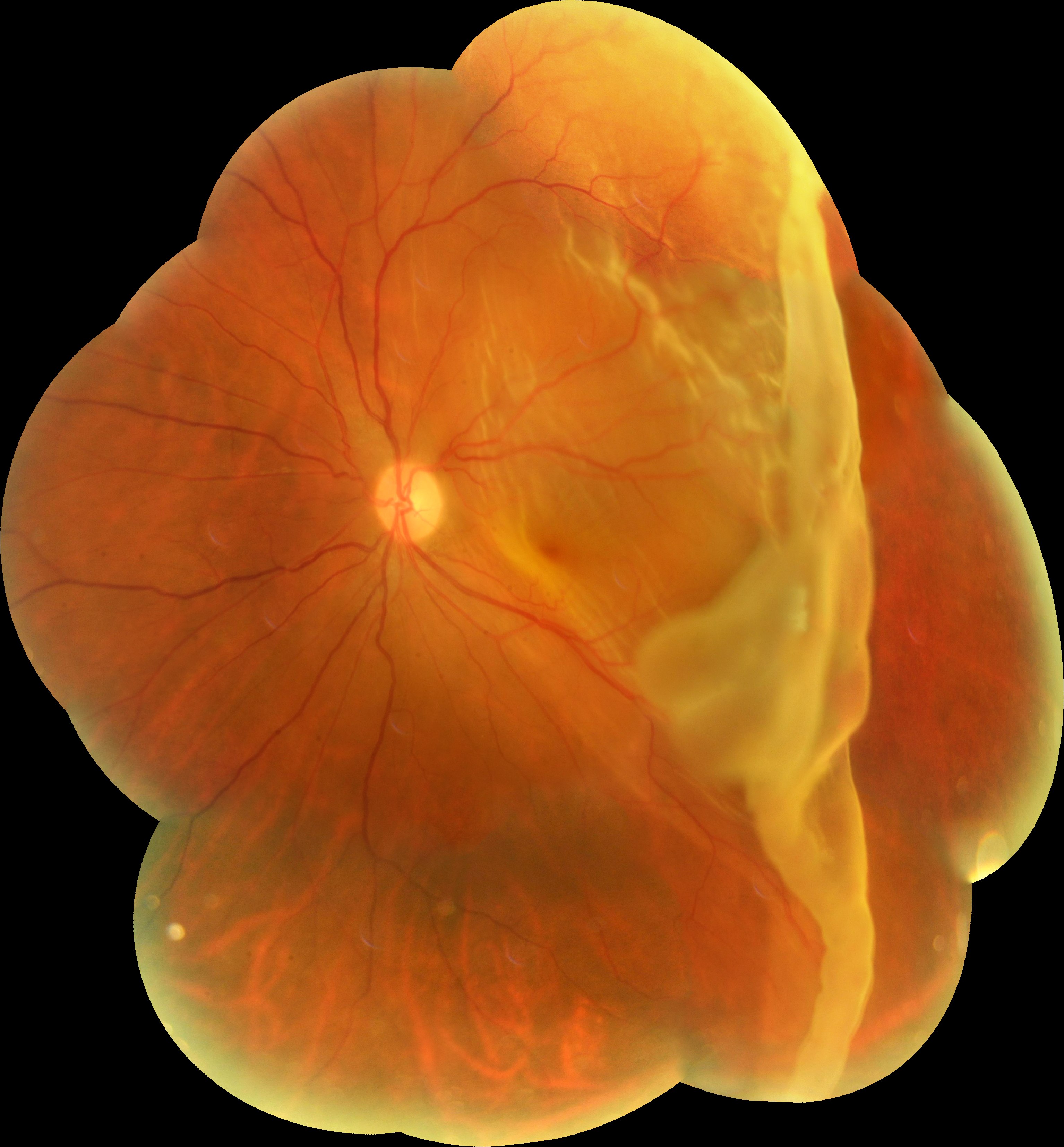

Prophylactic systemic antibiotics should be administered to patients to prevent endophthalmitis after a mechanical globe rupture or laceration. Patients with a suspected mechanical globe injury should have a metal shield placed over the eye, be given antiemetics, and be referred immediately to an ophthalmologist for surgical repair. Systematic review evaluating four heterogeneous clinical trials 1Ī chemical eye injury should be irrigated with normal saline or lactated Ringer solution until the ocular surface pH has normalized. Patients with a central retinal artery occlusion should receive a workup for stroke consisting of echocardiography, electrocardiography, and carotid artery ultrasonography. Patients with symptomatic floaters and flashing lights should be referred to an ophthalmologist for a dilated funduscopic examination to evaluate for a retinal tear or detachment. The eye should be covered with a metal shield until evaluation by an ophthalmologist. Physicians should administer prophylactic oral antibiotics after a globe injury to prevent endophthalmitis. A globe laceration or rupture is common in patients with a recent history of trauma from a blunt or penetrating object. Chemical injuries require immediate irrigation of the eye to neutralize the pH of the ocular surface. Patients with a central retinal artery occlusion require urgent referral for stroke evaluation and should receive therapy to lower intraocular pressure and vasodilating agents to minimize retinal ischemia. A detached retina is a serious problem that can. When detachment occurs, vision is blurred. Family physicians should be able to recognize the signs and symptoms of each condition and be able to perform a basic eye examination. The retina sends visual images to the brain through the optic nerve. Patients come in the morning, get the surgery done in the Yale Eye Center surgery center or a hospital, and then leave the hospital or the surgery center by noon.Central retinal artery occlusions, chemical injuries, mechanical globe injuries, and retinal detachments are eye emergencies that can result in permanent vision loss if not treated urgently.

Retinal surgery is usually done in an outpatient basis. “Some testing is done to evaluate the level of the condition.” “The physician needs to make sure that there is a good reason to do the surgery,” Dr. Pneumatic retinopexy, when a gas bubble is injected into the vitreous space inside the eye, pushing the retinal tear into place against the back wall of the eye via cryotherapy or laser surgery.

Vitrectomy, in which the vitreous gel is removed and replaced with a gas bubble or oil bubble to hold the retina in place.Scleral buckle, in which a flexible band is placed around the eye to counteract the force pulling the retina out of place.Retinal detachment surgery options include: An ophthalmologist may also suggest cryotherapy, which freezes the retina around the retinal tear and creates a scar that helps to hold the retina in place on the eye wall.Ī detached retina will require surgery. “It fixes the tear so it prevents further retinal detachment happening,” explains Dr. The ophthalmologist may suggest a laser treatment, which is very effective for retinal tears. The process for retinal surgery begins with a clinical evaluation and consultation. If the problem is located at the center of the retina (called the macula) the central field of vision will seem to be blurry. “Retinal detachment is like a curtain that comes from one side, and it slowly expands,” says Ron Adelman, MD, director of Yale Medicine's Retina & Vitreous Program. Fluid may pass through a retinal tear, lifting the retina off the back of the eye-much like wallpaper can peel off a wall. Sometimes the vitreous pulls hard enough to tear the retina in one or more places. (Think of it as the film detaching from the camera.) Once a retinal tear occurs, that vitreous gel-like fluid may seep through and lift the retina off the back wall of the eye, causing the retina to detach or pull away. If it takes a piece of the retina with it, you have a retinal tear. Sometimes inflammation or age-related nearsightedness can cause this gel to pull away. The eyeball is filled with vitreous gel, a clear substance that is attached to the retina. If you think of the eye as a camera, the lens is in the front, while the retina acts as the film.


 0 kommentar(er)
